Evolution of the Eye by Tony Thorp
Evolution of the eye
At the last meeting 19th oct Tony Thorp gave the second of his very interesting talks on the Evolution of the Eye.
To begin he gave a summary of the previous talk which had taken the time-line of evolution back to the late Cambrian.
Eyesight began with light sensitive cells which could detect light but not direction, images or shapes. The next step was the development of pits covered in photoreceptors. These were able to detect direction of light but not images or shapes. This allowed development of phototropism. Further evolutionary change led to deepening of the pit and the cavity becoming fluid filled and eventually covered over. The narrowing of the aperture produced the “pinhole camera eyes” as in Nautilus. This increased directional detection and some shape detection but not colour or depth.
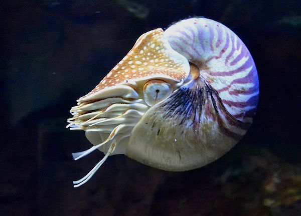
Nautilus by Archibald Tuttle Creative Commons https://creativecommons.org/licenses/by-sa/4.0/deed.en
Eventually the eye developed a lens so that now there was a fluid filled eye with lens and cornea which allowed image focusing which was followed by the development of the hard lens we know today which can focus light much more accurately. Tony concluded the summary of the first talk by talking about lamprey vision. The lamprey split off from the rest of the vertebrate group about 500MA. What is of interest is the rods and cones of the lampreys utilise the same mechanism of photo-transduction as other vertebrates therefore suggesting the mechanisms used in vision emerged very early on.
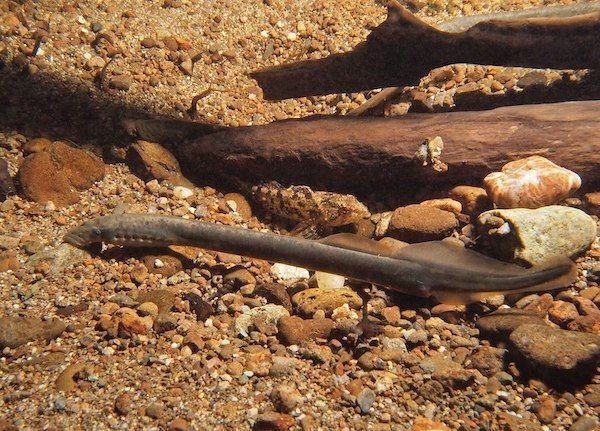
Western Brook lamprey bu US Fish and Wildlife Service Creative Commons
https://en.wikipedia.org/wiki/en:Creative_Commons
In the second half of his talk Tony showed that the development of vision could be shown to have evolved even further back than the Late Cambrian.
In order to understand evolution of vision we first have to understand the biochemical pathway of photo-transduction. The pathway involves a cGMP (cyclic guanosine mono-phosphate) - gated mechanism which alters the flow of ions across the cell membrane. Each photo-pigment molecule consists of the trans-membrane G -receptor protein opsin and a chromophore 11 cis -retinal, the whole known as rhodopsin. When light is absorbed the rhodopsin becomes enzymatically active which causes a cascade of events which culminates in the decrease in cGMP concentration which causes closure of the cGMP - gated channels on the plasma membrane. Sodium and calcium channels in the plasma membrane of the outer segment are kept open in the dark by a high level of cGMP. A decrease in cGMP leads to a decrease in influx of the cations into the cell and therefore hyperpolarization of the cell membrane. This neuronal signal is then transmitted to the brain.
Research has shown that there are two classes of opsin. Type I opsin, common in bacteria, evolved separately from type II opsin that are common in animals. Opsin is a member of large family of detector proteins, called the 'G-protein coupled receptors' (GPCRs).
Further, all known visual pigments in Neuralia ( the clade containing Cnidaria,eg. Corals and jellyfish; Ctenophora- Comb Jelly; and Bilateria -animals with bilateral symmetry) are composed of an opsin and a light-sensitive chromophore, usually retinal. Also these opsins can be classified into the same three subfamilies into which the bilaterian opsins are classified: the ciliary, rhabdomeric, and go-coupled plus retinochrome, retinal G protein-coupled receptor (Go/RGR) opsins.
It has been found that opsins only evolved after sponges had diverged from other animals, but before the split between Bilateria and Cnidaria. Within this time there is one animal lineage i.e the Placozoans which are very simple organisms and do contain opsin although cannot detect light.
Therefore it is thought that opsin itself evolved at some point between 755-711MA with it’s ability to detect light evolving somewhere between 711 and 700MA